Virtual Library | Cataracts and Cataract Surgery

If your pet is exhibiting signs of eye redness, swelling, discharge, cloudiness, squinting or bumping into objects around the house, it’s time to have their eyes examined for cataracts. Cataracts are a leading cause of vision impairment in animals. Read on to find out more about surgical solutions and related conditions associated with this disease.

← Flip through our digital brochure

A cataract is an opacity or cloudiness that develops within the lens of the eye. The lens is a normally clear structure in the center of the eye that helps to focus light on the retina and allow for fine detail in vision. Therefore, when a cataract develops, it results in cloudy or blurred vision, and up to complete loss of vision.
Cataracts are a leading cause of vision impairment in animals.

Most cataracts in dogs form due to genetics (inherited cataracts). Cataracts can also result from systemic disease (diabetes mellitus), inflammation within the eye (uveitis), trauma, toxic or nutritional causes, and with advanced age (senile cataracts). Genetic cataracts are more common in certain breeds of dogs, including Boston Terriers, Poodles, American Cocker Spaniels, Schnauzers, and Siberian Huskies, as well as Labrador, Chesapeake Bay and Golden Retrievers, among others. 

Cataracts may progress slowly or rapidly, depending on the underlying cause, age, and breed of dog. For example, an older Poodle or Boston Terrier will tend to have more slowly progressive cataract formation, while a younger Siberian Husky, Schnauzer, or Bichon Frise will tend to have more rapidly progressive cataracts that can cause significant inflammation within the eye and lead to secondary complications such as retinal detachment. Dogs with diabetes can develop blinding cataracts literally overnight. 

The only true form of cataract treatment is removal of the cataract with surgery. Use of topical anti-inflammatory drops can reduce complications secondary to the cataract, but will not decrease the cataract present. Other drops are touted to dissolve cataracts, however evidence is still lacking for any topical formulation. Aldose reductase inhibitors show promise in the future. At present, the only treatment is complete surgical removal.

If your pet has been diagnosed with cataracts, have an evaluation performed by a board-certified veterinary ophthalmologist. They will assess the severity, predisposition to secondary complications, and presence of concurrent ocular disease such as corneal disease, severe inflammation within the eyes (uveitis), or retinal disease (degeneration or detachment). 

If the ocular exam results determine your pet is an acceptable candidate, functional testing of the retina (electroretinogram) and ocular ultrasound will be performed to more definitively assess the potential of restoration of vision with surgery. 

Prior to surgery, your pet’s general health will be assessed with bloodwork and a physical examination to ensure he or she is a reasonable anesthetic candidate. Even aged pets can be considered reasonable anesthetic and cataract patients and restoration of vision can greatly enhance their quality of life.

The success rate for cataract surgery in dogs is quite high, with greater than 90% of cases undergoing a successful procedure and having improved vision following surgery.

This rate is mildly reduced in some instances, such as hypermature (chronic) cataracts, and dramatically reduced in other instances, such as lens rupture within the eye (phacoclastic uveitis).

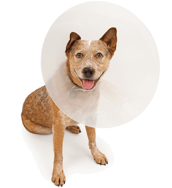
Your pet will need to wear an Elizabethan collar or “cone” to prevent inadvertent trauma to the surgical site for the first 7-10 days following surgery.

The main complications following cataract surgery include glaucoma (increased pressure in the eye), eye infection (endophthalmitis), retinal detachment or corneal ulceration. 

Glaucoma occurs in roughly 5% of cases following surgery and is typically manageable with eye drops to control the pressure. Rarely, eye drops are ineffective alone to control the pressure, and an additional procedure (laser surgery) will be recommended. 

The development of both an eye infection or retinal detachment can be severe, vision-threatening complications. However, every precaution is taken to avoid these outcomes, and fortunately occurs in less than 1% of cases.

Cataract surgery in dogs is very similar to cataract surgery in people. 

  • A small incision is made at the top part of the eye and a circular hole is created within the outer flexible capsule of the lens. 
  • Ultrasound energy (phacoemulsification) is used to remove the internal cloudy lens material. 
  • The capsule is polished, an artificial lens is implanted within it, and the incision at the top of the eye is closed with a suture.
  • In rare cases of capsule instability, a replacement lens is either not implanted, or is sutured within the eye. Without a lens implant, your pet will still have improved vision, but will be farsighted.

Plan to bring your pet for an exam roughly 3-4 times in the first month following surgery. The most common recheck schedule is 1-day, 1-week, 2-weeks, and 1-month following surgery. This rigorous routine provides the best chance of detecting potential complications at an early stage when treatment interventions are more effective. Long-term follow up appointments typically occur at 3 months,
6 months, 1 year, and then annually as needed.

At Animal Vision Center, we want the procedure to be a success for your pet just as much as you do. All recheck evaluations during the first month are included in the cost of the surgery, and we ask that you call or bring in your pet for evaluation if there is any question as to how he or she is doing at home.