Virtual Library | Glaucoma & Glaucoma Surgery
Glaucoma is one of the leading causes of blindness in both animals and people. Without aggressive treatment, glaucoma can lead to irreversible damage to the optic nerve and permanent loss of vision. Read on to learn more about the signs, causes and treatment options.
← Flip through our digital brochure
PLEASE NOTE: It is important to remember that successful treatment of glaucoma requires early diagnosis, rapid treatment and long-term vigilant monitoring under the care of a veterinary ophthalmologist.
Visit related pages for more information:
 Glaucoma is a condition in which inadequate drainage of fluid from the eye results in increased pressure within the eye and damage to the optic nerve. It is one of the leading causes of blindness in both animals and people. There are many different types of glaucoma and reasons why it develops. Without aggressive treatment, glaucoma can lead to irreversible damage to the optic nerve and permanent loss of vision.
 The eye is a fluid-filled structure. This fluid (aqueous humor) is produced by the ciliary body within the eye and flows throughout the eye to nourish the structures within it. The fluid is produced at a relatively constant rate and normally flows out of the eye through drainage holes that circle around the eye between the iris and cornea (the iridocorneal angle). When there are abnormalities in the drainage of fluid from the eye, pressure elevates, leading to the development of glaucoma.
Glaucoma can occur due to a primary, inherited abnormality in the drainage angle, or secondary to other problems within the eye. Primary glaucoma is common in many purebred dogs, but can also occur in mixed breed dogs. Secondary glaucoma occurs due to inflammation within the eye, shifting of the lens out of position (lens luxation), cataract formation, trauma, retinal detachment and intraocular tumors/cancer, among other causes.
Acute development of glaucoma is a medical emergency and must be treated immediately, or permanent vision loss can occur. It can be treated medically or surgically. Medical therapy for glaucoma is generally very effective in the early stages, however with time and continued changes in the eye, medical therapy generally fails. Surgical therapy is the best option for potential long-term control of the pressure and maintenance of comfort and vision.
MEDICAL THERAPY:
 There are numerous medications used to help control glaucoma within the animal eye, including:
- Carbonic anhydrase inhibitors (dorzolamide, brinzolamide, methazolamide)
- Prostaglandin analogues (latanoprost, travoprost, bimatoprost)
- Beta blockers (timolol)
- And occasionally parasympathomimetics (demecarium bromide, pilocarpine)
These medications are often combined with pain medications, anti-inflammatory therapy and medications to help prevent continued damage of the retina and optic nerve after control of the pressure. As stated above, medical therapy is commonly effective in the short term, but generally fails in the long run, requiring higher and higher dosages of medications until the pressure is no longer manageable and vision loss occurs.
SURGICAL THERAPY TO PRESERVE VISION:
Surgical therapy to control glaucoma consists of procedures that increase the flow of fluid out of the eye (shunt procedures) and procedures to decrease the amount of fluid produced in the eye (laser surgery). Shunt procedures do not tend to have a long-term benefit alone, but are occasionally combined with laser surgery for long-term control. There are several types of laser surgery that can be performed, including micropulse laser therapy, transscleral laser surgery or endoscopic laser surgery. Your veterinary ophthalmologist will determine which laser therapy is best for your pet.
END-STAGE THERAPY:
When blindness occurs secondary to glaucoma and the pressure is uncontrolled, there are several end-stage procedures that can be considered to provide comfort.
- The first procedure involves performing a painless injection into the back of the eye to lower the production of fluid within the eye. This procedure is effective approximately 85% of the time but can result in a decrease in the size of the globe, cataract formation and cloudy appearance to the eye.
- The last two procedures are surgical in nature and require general anesthesia. Enucleation is the complete removal of the globe and placement of a prosthetic within the orbit with the eyelids permanently closed. This procedure results in the most rapid control of the pain and the lowest potential for possible complications. Evisceration and intraocular prosthesis placement involves removing the internal contents of the globe and placing the prosthetic on the inside of the eye to result in a more normal post-operative appearance of an eye, but leaves the potential for problems with the surface of the eye such as ulcer formation or keratitis (inflammation of the cornea).
The acute clinical signs of glaucoma generally include a cloudy eye with a red, bloodshot appearance; tearing, or ocular discharge; and pain (squinting). Due to the pain, some animals will also act more tired than usual, have a poor appetite and exhibit nausea or vomiting. Over time, without treatment, the eye will become swollen and larger in appearance and will lose vision permanently.
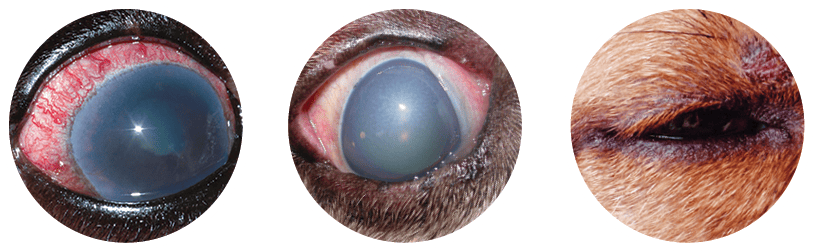 COMMON SIGNS OF GLAUCOMA: CLOUDY EYE WITH BLOODSHOT APPEARANCE, TEARING OR OCULAR DISCHARGE, AND SQUINTING/PAIN.
 Breeds with the highest incidence of primary inherited glaucoma include the American Cocker Spaniel, Basset Hound, Chow Chow, Shar Pei and Boston Terrier.
Other breeds which are predisposed to glaucoma include many terrier breeds (Cairn, Fox, Jack Russell), poodle breeds (toy, miniature, standard), arctic breeds (Samoyed, Siberian Husky, Akita), spaniel breeds (English Cocker, English/Welsh Springer, Brittany), as well as other breeds such as the Beagle, Bichon Frise, Bouvier, Shih Tzu, Llasa Apso, and Pekingese, among others. Breeds predisposed to inherited luxation of the lens and secondary glaucoma include the Jack Russell Terrier, Fox Terrier (and other terrier breeds) and the Shar Pei.
So, You Brought Home a Bulldog
Cataracts and Cataract Surgery
Entropion Causes and Treatment
Feline Herpesvirus and Treatment
OFA Certification Registry Exams
Visit our blog, In Focus, to learn more about the pets we see, the treatments we offer and the services we provide to help your pet “see a better life.”