Surgical therapies for the management of KCS include the cyclosporine implant, parotid duct transposition and in very rare circumstances, enucleation, or removal of the eye.
Placement of a cyclosporine implant can result in significant improvement in tear production and reduction in ocular surface inflammation for approximately one year following surgery. Improvement can occur in both cases that respond to therapy with topical cyclosporine or tacrolimus, and those that do not. This procedure may need to be repeated on a long-term basis.
The parotid duct transposition works by re-routing a salivary duct from the mouth to the eye. This results in lubrication of the ocular surface with saliva. While exceptionally helpful in select cases, this surgery is generally reserved for refractory cases since it can result in the development of other ocular surface problems such as mineral formation (similar to plaque buildup on teeth); potential for skin infection under the eye due to less control over the secretion of saliva than tears; and risk of duct damage during the translocation to the eye.
The last surgical procedure that is infrequently considered for severe KCS is enucleation, or removal of the eye. This surgery is reserved for chronically painful eyes that have become blind from scarring of the surface or severe corneal ulceration. This procedure can provide significant return of comfort and quality of life in animals suffering with severe forms of KCS.
 Dry Eye, also known as Keratoconjunctivitis sicca (KCS), is a condition in which a lack of tear production leads to ocular surface inflammation, discomfort and increased rates of secondary complications such as corneal ulceration and scarring of the ocular surface. There are many and varied causes of KCS, but it most commonly occurs as a result of genetically-based autoimmune inflammation within the tear-producing glands of the eye. Once the underlying cause is identified, appropriate therapy can be administered.
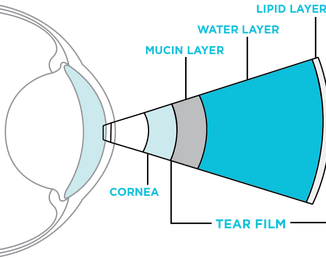
 The eye is a delicate structure and the first layer of protection is the ocular tear film, a complex structure made of multiple layers of interacting elements. This water-based layer of the tear film serves many functions including optics; lubrication and clearing of debris from the ocular surface; nourishment of the ocular surface; and antimicrobial and immune functions. When this important layer of protection is missing, the ocular health suffers.
 Breeds with the highest incidence of KCS include the English bulldog, West Highland White terrier, Lhasa Apso, Pug, Pekingese, American cocker spaniel, Yorkshire terrier and Shih tzu.