Virtual Library | Corneal Ulcers and Treatment
The term ulcer indicates loss of corneal tissue resulting in an open wound on the surface of the eye. Ulcers can be classified as superficial or deep, depending on the amount of tissue that is lost. Such loss can involve just a few cell layers and heal very quickly, or it can affect the entire depth of the cornea, taking weeks or months to fully heal. Read on to learn more about corneal types and treatment.
← Flip through our digital brochure
 The cornea is the clear dome on the surface of the eye. It is the major refractive surface of the eye, and problems in the cornea may lead to decreased vision. The cornea is covered by an outer protective tear film which is crucial for ocular surface health. It also has a robust nerve supply; therefore corneal problems can be very painful. The cornea is composed of three layers: epithelium, stroma, and endothelium (including Descemet’s membrane).
 The term ulcer indicates loss of corneal tissue resulting in an open wound on the surface of the eye. Ulcers can be classified as superficial or deep, depending on the amount of tissue that is lost. Such loss can involve just a few cell layers and heal very quickly, or it can affect the entire depth of the cornea, taking weeks or months to fully heal.
The cause of a corneal ulcer is not always apparent. Most commonly, ulcers form due to irregularities of the ocular surface or trauma. Ocular surface irregularities can include overly long or irregular eyelids, eyelashes rubbing on the cornea, tear deficiency, decreased corneal sensitivity/nerve deficiency or degeneration of the cornea. Trauma to the ocular surface typically occurs when a foreign object (another pet’s claw, grass blade, sand) or a chemical irritant (cleaning products, etc.) comes in contact with the cornea.
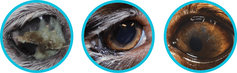
Common causes of corneal ulcers: Dry Eye (tear deficiency), Entropion (rolling in of eyelid margin) and Distichia (abnormal eyelid hair).
Breeds with prominent eyes and short noses, or brachycephalic breeds, have an increased rate of corneal ulcer formation due to a combination of increased exposure of their eyes, decreased tear production and decreased corneal sensation. This combination of traits can quickly allow a simple ulcer to progress rapidly into a deep, perforating ulcer, requiring urgent therapy. Brachycephalic breeds most predisposed to corneal ulcer formation include the Shih tzu, Pekingese, Pug, Boston Terrier, Boxer, French Bulldog and English Bulldog. There is also an increased rate of ulcer formation in animals with underlying endocrine disorders such as diabetes mellitus, hypothyroidism and Cushing’s syndrome, as well as in animals with underlying allergies and ear infections.
 SIMPLE
A simple ulcer forms due to a minor abrasion of the cornea. These ulcers generally heal within a few days, often before a problem is even noticed.
.

IRRITANT-ASSOCIATED
Treatment of an ulcer that has formed as the result of an irritant, such as an embedded grass awn, abnormal eyelashes or an eyelid mass rubbing on the surface of the eye, requires elimination of the irritant for the ulcer to heal. Surgery is often required to correct the problem.
..

INFECTED
Any type of ulcer can become infected with bacteria. Specialized tests will help to determine the type of infection present. An infected ulcer requires the use of targeted, aggressive antibiotic therapy, or in some cases, surgery to stabilize the corneal surface. Infected ulcers can progress through the cornea rapidly. When the ulcer reaches 50% depth in the cornea, surgical stabilization with a corneal or conjunctival graft is generally recommended. Your family veterinarian or a veterinary ophthalmologist will make this assessment to help prevent rupture and potential loss of your pet’s eye.

INDOLENT
An indolent ulcer forms in an older dog due to a breakdown in the connections between the top layers of the cornea. This type of ulcer requires treatment with a procedure called a grid keratotomy to allow for complete healing. Your family veterinarian may refer you to a veterinary ophthalmologist for this procedure.
Conjunctival tissue is the thin tissue containing blood vessels that surrounds the eye. During surgery, this tissue and/or nearby healthy corneal tissue is used to repair the defect in the cornea. It is secured in place with very fine suture material that is ultimately absorbed by the body and does not need to be removed. This tissue strengthens the weakened region and helps to prevent rupture of the eye.

Following surgery, your pet will remain on medical therapy to prevent infection, decrease inflammation and maintain comfort. An Elizabethan collar (plastic cone) will be required for the first week to prevent self-trauma to the surgery site. The cornea will remain cloudy for the first few weeks following surgery and will become clearer again 1-2 months following surgery.
At Animal Vision Center, we want the procedure to be a success for your pet just as much as you do. We ask that you call or bring in your pet for evaluation if there is any question as to how he or she is doing at home.
So, You Brought Home a Bulldog
Cataracts and Cataract Surgery
Entropion Causes and Treatment
Feline Herpesvirus and Treatment
OFA Certification Registry Exams
Visit our blog, In Focus, to learn more about the pets we see, the treatments we offer and the services we provide to help your pet “see a better life.”