Virtual Library | Prolapsed Nictitans Gland and Replacement
If you notice a red or pink mass protruding from behind your pet’s third eyelid, it’s time to have their eyes examined for a prolapsed nictitans gland, also known as “cherry eye.” Once the gland of the third eyelid has prolapsed, surgery is required to replace the gland. Read on to find out more about predispositions, causes and surgical solutions for a prolapsed nictitans gland.
← Flip through our digital brochure
So, You Brought Home a Bulldog
Cataracts and Cataract Surgery
Entropion Causes and Treatment
Feline Herpesvirus and Treatment
OFA Certification Registry Exams
Visit our blog, In Focus, to learn more about the pets we see, the treatments we offer and the services we provide to help your pet “see a better life.”
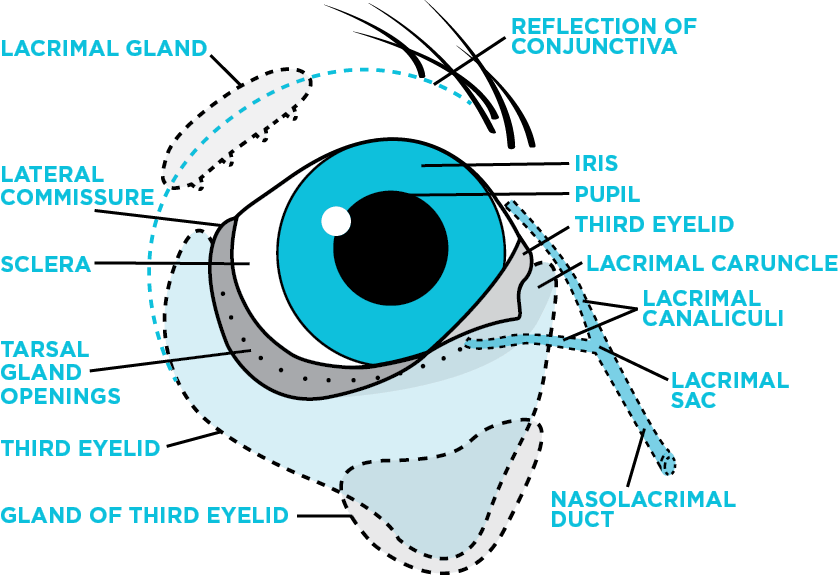
 Prolapsed nictitans gland, also known as “cherry eye,” occurs when the gland of the nictitans comes out of position. It protrudes from behind the third eyelid appearing as a red or pink mass. This appearance is how it gained its common name. This prolapsed gland can occur in one or both eyes. It can also regress then reappear, but more commonly the prolapsed gland remains exposed and out of position.
 The third eyelid, also known as the nictitating membrane or nictitans, is a membrane that sits just inside of the lower eyelid in dogs and cats. The nictitans contains a gland that typically sits at the base of this membrane. This gland plays an important role in maintaining normal tear production and is responsible for 40-60% of the tears. Damage to this gland can lead to low-tear production. Normal tear production is important to maintain ocular surface health. The third eyelid also contains cartilage for structural support and immune tissue to provide antibodies in the tear film.
 Once the gland of the third eyelid has prolapsed, surgery is required to replace the gland.
 Dry eye is uncomfortable
 Some breeds of dogs and cats have a conformational predisposition for a weaker supportive ligament. Brachycephalic breeds (those with prominent eyes and short noses) are more often affected due to their stout facial conformation. Among these, those that are more predisposed to nictitans gland prolapse include: the Shih Tzu, Lhasa Apso, English Bulldog, Boston Terrier and Burmese cats. Other dog breeds that are predisposed to this condition include the Cocker Spaniel, Cavalier King Charles Spaniel, Beagle, Poodle, Bloodhound and Mastiff, although it can be seen in any young pet.
 To replace the gland, the surgeon will create a pocket to help position the gland back into its normal location. They will make a curved incision on both sides of the gland where it is prolapsed. The outer edges of these incisions are then brought together to form a pocket using very fine suture material that is ultimately absorbed by the body. This pocket covers, protects and replaces the previously exposed gland.
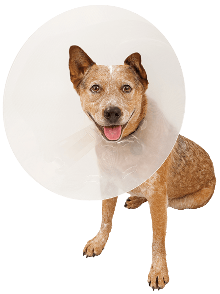
 Following surgery the pet patient will start medical therapy to prevent infection, decrease inflammation and maintain comfort. An Elizabethan collar (plastic cone) is required for the first two weeks to prevent self-trauma to the surgery site. The third eyelid will have mild to moderate redness and swelling immediately following surgery, but this will continue to resolve over the first few weeks.